古病理学
Paleopathology
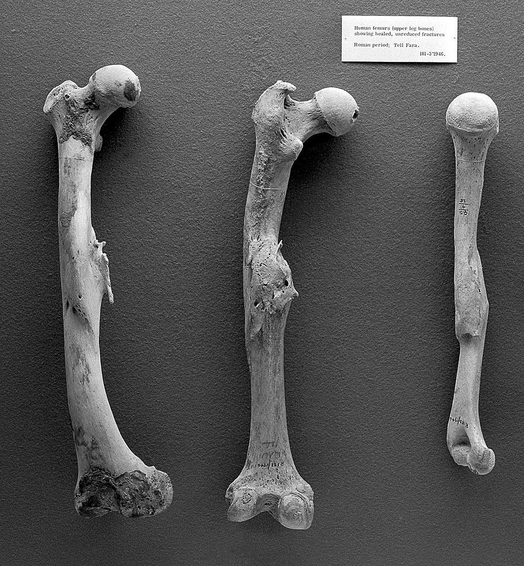
ローマ時代のヒトの大腿骨と上腕骨(右)
★古病理学[Paleopathology]は、古病理学とも表記さ れ、化石、ミイラ化した組織、骨格、コプロライトの分析などの検査を通して、生物の古代の病気や傷害を研究することである。古代の人間の病気を研究する際 の 具体的な資料としては、初期の文書、初期の書籍の挿絵、過去の絵画や彫刻などが考えられる。"Paleopathology "という言葉の個々の語源を見ると、それが何を包含しているのかの基本的な定義ができる。"Paleo "は「古代の、初期の、先史時代の、原始の、化石」を意味する。また、"-pathology "という接尾辞は、ラテン語で「病気の研究」を意味するpathologiaに由来する[1]。上記のものを分析することで、病気の進化や過去の文明の 治療方法に関する情報は、いずれも貴重な副産物となる。歴史的にヒトを対象とした研究が行われてきましたが、ヒトが他のどの動物よりも病気になりやす いという証拠はない[2]。
現在、中止されている先住民の骨の研究を再会するのに「日本人の祖先」や「未来の皆さんの病
気解明になる」というリップサービスを人類学者はやめよ!!それらを古病理学者や司法人類学者が真摯に研究すれば栄養失調や衛生状態という過去の政治の瑕
疵が百出するだろう——垂水源之介(2023)
| Paleopathology,
also spelled palaeopathology, is the study of ancient diseases and
injuries in organisms through the examination of fossils, mummified
tissue, skeletal remains, and analysis of coprolites. Specific sources
in the study of ancient human diseases may include early documents,
illustrations from early books, painting and sculpture from the past.
Looking at the individual roots of the word "Paleopathology" can give a
basic definition of what it encompasses. "Paleo-" refers to "ancient,
early, prehistoric, primitive, fossil." The suffix "-pathology" comes
from the Latin pathologia meaning "study of disease."[1] Through the
analysis of the aforementioned things, information on the evolution of
diseases as well as how past civilizations treated conditions are both
valuable byproducts. Studies have historically focused on humans, but
there is no evidence that humans are more prone to pathologies than any
other animal.[2] Paleopathology is an interdisciplinary science, meaning it involves knowledge from many sectors including (but not limited to) "clinical pathology, human osteology, epidemiology, social anthropology, and archaeology".[3] It is unlikely that one person can be fluent in all necessary sciences. Therefore, those trained in each are important and make up a collective study. Training in Anthropology/archaeology are arguably most important because the analysis of human remains and ancient artifacts are paramount to the discovery of early disease. |
古病理学[Paleopathology]は、古病理学とも表記さ
れ、化石、ミイラ化した組織、骨格、コプロライトの分析などの検査を通して、生物の古代の病気や傷害を研究することである。古代の人間の病気を研究する際
の
具体的な資料としては、初期の文書、初期の書籍の挿絵、過去の絵画や彫刻などが考えられる。"Paleopathology
"という言葉の個々の語源を見ると、それが何を包含しているのかの基本的な定義ができる。"Paleo
"は「古代の、初期の、先史時代の、原始の、化石」を意味する。また、"-pathology
"という接尾辞は、ラテン語で「病気の研究」を意味するpathologiaに由来する[1]。上記のものを分析することで、病気の進化や過去の文明の
治療方法に関する情報は、いずれも貴重な副産物となる。歴史的にヒトを対象とした研究が行われてきましたが、ヒトが他のどの動物よりも病気になりやす
いという証拠はない[2]。 古病理学は学際的な科学であり、「臨床病理学、人骨学、疫学、社会人類学、考古学」を含む(ただしこれらに限定されない)多くの分野からの知識を含んでい ます[3]。したがって、それぞれの訓練を受けた者が重要であり、集合的な研究を構成している。人類学や考古学の訓練は、間違いなく最も重要です。なぜな ら、人間の遺体や古代の遺物を分析することは、初期の病気を発見するために最も重要だからです。 |
| History Historical evidence shows that deviations from good health have long been an interest to humans. Although the content that makes up this study can be traced through ancient texts, the term "paelopathology" did not have much traction until the 20th century. This time period saw an increase in case studies and "published reports on ancient diseases".[4] Ancient texts that are thousands of years old record instances of diseases such as leprosy. From the Renaissance to the mid-nineteenth century, there was increasing reference to ancient disease, initially within prehistoric animals although later the importance of studying the antiquity of human disease began to be emphasized. Some historians and anthropologists theorize that "Johann Friederich Esper, a German naturalist...heralds the birth of paleopathology."[5] Although it wasn't until between the mid nineteenth century and World War I that the field of human paleopathology is generally considered to have come about. During this period, a number of pioneering physicians and anthropologists, such as Marc Armand Ruffer, G. Elliot Smith, Frederic Wood Jones, Douglas E. Derry, and Samuel George Shattock, clarified the medical nature of ancient skeletal pathologies.[6] This work was consolidated between the world wars with methods such as radiology, histology and serology being applied more frequently, improving diagnosis and accuracy with the introduction of statistical analysis. It was at this point that paleopathology can truly be considered to have become a scientific discipline.[7] Today, the use of biomedical technology like DNA and isotopic analysis are major developments for pathological knowledge.[8] After World War II paleopathology began to be viewed in a different way: as an important tool for the understanding of past populations, and it was at this stage that the discipline began to be related to epidemiology and demography. New techniques in molecular biology also began to add new information to what was already known about ancient disease,[7] as it became possible to retrieve DNA from samples that were centuries or millennia old. |
歴史 健康状態からの逸脱は、古くから人間の関心事であったことを示す歴史的な証拠があります。この研究を構成する内容は古文書から辿ることができるが、「ペー ロパソロジー」という用語は20世紀まであまり普及していなかった。この時代には、ケーススタディや「古代の病気に関する公表された報告書」が増加した [4]。数千年前の古文書には、ハンセン病のような病気の事例が記録されている。 ルネサンスから19世紀半ばにかけて、古代の病気に関する言及が増え、最初は先史時代の動物が対象だったが、後に人間の病気の古さを研究することの重要性 が強調され始めた。歴史学者や人類学者の中には、「ドイツの博物学者ヨハン・フリードリヒ・エスパーが古病理学の誕生を告げた」とする説もある[5]が、 一般に人間の古病理学の分野が生まれたと考えられているのは、19世紀半ばから第一次世界大戦までの間である。この時期、マーク・アルマン・ラファー、 G・エリオット・スミス、フレデリック・ウッド・ジョーンズ、ダグラス・E・デリー、サミュエル・ジョージ・シャトックといった多くの先駆的医師や人類学 者が、古代骨格病理の医学的性質を明らかにした[6]。この仕事は世界大戦の間に統合され、放射線学、組織学、血清学といった方法がより頻繁に応用され、 統計分析の導入によって診断と正確さが向上した。今日、DNAや同位体分析などの生物医学的技術の利用は、病理学的知識にとって大きな進展となっている [8]。 第二次世界大戦後、古病理学は、過去の集団を理解するための重要なツールとして、別の見方をされるようになり、この段階から、疫学や人口統計学と関連する 学問となりました。 また、分子生物学の新しい技術により、数世紀あるいは数千年前のサンプルからDNAを取り出すことが可能になり、古代の病気についてすでに知られていたこ とに新たな情報が加わるようになった[7]。 |
| Methods and techniques To analyze human remains of the past, different techniques are used depending on the type of remains that are found. For example, "the approach to palaeopathological samples depends on the nature of the sample itself (e.g. bone, soft tissue or hair), its size (from minimal fragments to full bodies), the degree of preservation and, very importantly, the manipulation allowed (from intact sample ready for display to absolute access and freedom to undertake any kind of valuable destructive analysis including a full autopsy study)."[9] Much research done by archaeologists and paleopathologists is on bones. The basic nature of bones allows them to not degrade over time like other human remains would, making osteopathology important in studying ancient disease. Human osteopathology is classified into several general groups: Arthropathy Infection Oral pathology Trauma Tumor Whilst traumatic injuries such as broken and malformed bones can be easy to spot, evidence of other conditions, for example infectious diseases such as tuberculosis and syphilis, can also be found in bones. Arthropathies, that is joint diseases such as osteoarthritis and gout, are also not uncommon. The first exhaustive reference of human paleopathology evidence in skeletal tissue was published in 1976 by Ortner & Putschar.[10] In identifying pathologies, physical anthropologists rely heavily on good archaeological documentation regarding location, age of site and other environmental factors. These provide the foundation on which further analysis is built and are required for accurate populations studies. From there, the paleopathology researcher determines a number of key biological indicators on the specimen including age and sex. These provide a foundation for further analysis of bone material and evaluation of lesions or other anomalies identified. Archaeologists increasingly use paleopathology as an important main tool for understanding the lives of ancient peoples. For example, cranial deformation is evident in the skulls of the Maya, where a straight line between nose and forehead may have been preferred over an angle or slope. There is also evidence for trepanation, or drilling holes in the cranium, either singly or several times in a single individual. Partially or completely healed trepanations indicate that this procedure was often survived. The 10,000 year-old human remains discovered at the site of Nataruk in Turkana, Kenya, reportedly show extreme traumatic lesions to the head, neck, ribs, knees and hands, including embedded stone projectiles, and they may represent the earliest evidence of inter-group conflict between hunter-gatherers in the past.[11][12]  Evidence of skeletal trauma from violence Violence Differentiating skeletal trauma as the result of violence compared to that caused by accidental or other causes is achieved by integrating the skeletal analysis of mechanical injury to bone with the sociocultural context.[18] Intertwining the biological analysis with the sociocultural factors presented by not just the individual but also the larger group context has allowed bioarchaeology to identify numerous types of violence including, as The Routledge Handbook of Paleopathology notes,“warfare, ritualized combat, hand to hand fighting, raids and ransacking, massacres, torture, executions, witchcraft, captive taking, slavery, anthropophagy, intimate partner and child abuse, scalping and human sacrifice."[16] Without this synthesis of the biological analysis and social theory, Klaus notes that trauma studies are reduced to “simply descriptions of trauma found on bone.”[19] |
方法と技法 過去の人骨を分析するためには、発見された遺体の種類によって異なる技法が用いられます。例えば、「古病理学的サンプルへのアプローチは、サンプル自体の 性質(骨、軟組織、毛髪など)、サイズ(最小限の断片から全身まで)、保存の程度、そして非常に重要なのは、許される操作(展示可能な無傷のサンプルか ら、完全解剖調査を含むあらゆる種類の貴重な破壊的分析を実施できる絶対アクセスと自由まで)によって決まる」 [9] 考古学者と古病理学者の多くの研究は、骨について行われています。骨の基本的な性質により、他の人骨のように時間の経過とともに劣化することがないため、 古代の病気を研究する上で骨病理学は重要である。人間の骨病理学は、いくつかの一般的なグループに分類される: 関節症(Arthropathy 感染症 口腔病理学 外傷 腫瘍(しゅよう 骨折や奇形などの外傷はわかりやすいのですが、結核や梅毒などの感染症も骨から発見されることがあります。また、変形性関節症や痛風など、関節の病気も少 なくありません。 骨格組織におけるヒトの古病理学的証拠に関する最初の包括的な文献は、1976年にOrtner & Putscharによって出版されました[10]。病理の特定において、身体人類学者は、場所、場所の年代、その他の環境要因に関する優れた考古学的文書 に大きく依存しています。これらは、さらなる分析の土台となるもので、正確な集団研究に必要なものである。そこから、古病理学研究者は、年齢や性別など、 標本の主要な生物学的指標を決定していきます。これらは、骨材をさらに分析し、特定された病変やその他の異常を評価するための基礎となるものです。 考古学者は、古代人の生活を理解するための重要な主要ツールとして、古病理学を利用することが増えています。例えば、マヤの頭蓋骨には頭蓋の変形が見ら れ、鼻と額の間の直線は、角度や傾斜よりも好まれた可能性があります。また、トレパネーション(頭蓋に穴を開けること)が、一人の人間に一度だけ、あるい は数回行われた形跡もある。トレパネーションは、部分的または完全に治癒していることから、この処置はしばしば存続していたことがわかります。ケニアの トゥルカナのナタルク遺跡で発見された1万年前の人骨は、頭部、首、肋骨、膝、手に極度の外傷性病変があり、石の突起物が埋め込まれていたと報告されてお り、過去の狩猟採集民のグループ間紛争の最古の証拠となる可能性がある[11][12]。 暴力による骨格外傷の証拠 暴力 暴力による骨格外傷と、事故やその他の原因による外傷とを区別するには、骨への機械的損傷に関する骨格分析と社会文化的文脈とを統合する必要がある。 [18] 生物学的分析を、個人だけでなくより広範な集団の文脈によって提示される社会文化的要因と結びつけることで、生物考古学は数多くの暴力の形態を特定するこ とが可能となった。例えば、『The Routledge Handbook of Paleopathology』が指摘するように、「 戦争、儀式化された戦闘、白兵戦、襲撃と略奪、虐殺、拷問、処刑、魔術、捕虜の捕獲、奴隷制、人食、パートナーや子供への虐待、頭皮剥ぎ、人身供犠」と いった数多くの種類の暴力を特定することが可能になった。[16] クラウスは、生物学的分析と社会理論のこの統合がなければ、外傷研究は「単に骨に見られる外傷の記述」に還元されてしまうと指摘している。[19] |
| Archaeological infectious
diseases Several diseases are present in the archaeological record. Through archaeological evaluation these diseases can be identified and sometimes can explain the cause of death for certain individuals. Aside from looking at sex, age, etc. of a skeleton, a paleopathologist may analyze the condition of the bones to determine what sort of diseases the individual may have. The goal of a forensic anthropologist looking at the Paleopathology of certain diseases is to determine if the disease they are researching are still present over time, with the occurrence of certain events, or if this disease still exists today and why this disease may not exist today.[13] Some disease that are found based on changes in bone include Tuberculosis Leprosy Syphilis Apart from bones, molecular biology has also been used as a tool of paleopathology over the last few decades, as DNA can be recovered from human remains that are hundreds of years old. Since techniques such as PCR are highly sensitive to contamination, meticulous laboratory set-ups and protocols such as "suicide" PCR are necessary to ensure that false positive results from other materials in the laboratory do not occur. For example, the long-held assumption that bubonic plague was the cause of the Justinian plague and the Black Death has been strongly supported by finding Yersinia pestis DNA in mass graves,[14][15] whereas another proposed cause, anthrax, was not found.[14] |
考古学上の感染症 考古学的な記録には、いくつかの病気が存在します。考古学的な評価を通じて、これらの病気を特定することができ、時には特定の個人の死因を説明することが できます。骨格の性別や年齢などを調べる以外にも、古生物学者は骨の状態を分析し、その個人がどのような病気にかかっていたかを判断することがあります。 特定の病気の古病理学を研究する法人類学者の目的は、研究している病気が、特定の出来事の発生によって、時を経ても存在するかどうか、あるいはこの病気が 今日でも存在するかどうか、なぜこの病気が今日存在しないかもしれないかを判断することです[13] 骨の変化に基づいて発見されるいくつかの病気は以下の通りです。 結核 ハンセン病 梅毒 骨以外にも、分子生物学はここ数十年、古病理学のツールとして利用されています。なぜなら、数百年前の人骨からDNAを回収することができるからです。 PCRのような技術は汚染に対して非常に敏感であるため、実験室内の他の物質による偽陽性を起こさないようにするためには、細心の注意を払った実験室の セットアップや「自殺」PCRのようなプロトコルが必要です。 例えば、ユスティニアヌス疫と黒死病の原因は、集団墓地からエルシニア・ペスティスのDNAが発見されたことにより、長年信じられてきた仮説が強く支持さ れた[14][15]が、別の原因と考えられていた炭疽菌は発見されなかった[14]。 |
Black Death The Black Plague, Florence 1348 The Black Death pandemic occurred between 1347 and 1351.[16] It is believed that the origin of the Black Death was bubonic plague.[16] Symptoms of Bubonic Plague include swollen lymph nodes, fever, headache, fatigue, and muscle aches.[17] Some people also developed swellings in which blood and pus seeped from them during the Black Death.[18] The Black Death originated in China and spread along trade routes and ports affecting many countries including North Africa and many European countries such as Italy, Spain, France, Germany, Switzerland, and Hungary.[16] It is estimated that the Black Death killed up to 200 million people.[19] In 2013, during an excavation in Thornton Abbey in North Lincolnshire, a mass grave was discovered with 48 people in it including 27 children.[20] Radiocarbon dating and artifacts found in the mass grave allowed archaeologists to narrow down the time period in which the bodies were from to the time of the Black Death.[20] The wide range of ages of the remains, from one to 45 years of age, led archaeologists to determine that something devastating most likely caused their deaths.[20] Typically, mass graves contain remains from either very young ages or very old ages: this was not the case here. Because all ages were being buried here, archaeologists could deduce that although Lincolnshire is a small town, it was consumed by the plague to the extent in which a mass grave was needed. A mass grave was up until this discovery, very rare because small towns seemed to bury their dead in usual ways.[21] It is believed that the reason for mass burials in Europe during this time were because of the overwhelming number of deaths caused by the Black Plague.[19] Archaeologists extracted teeth samples from the remains of the individuals who were buried, and it was revealed that there was the presence of plague bacteria.[20] These samples showed the presence of Y. pestis DNA, the bacterial cause of the plague. "Molecular identification by 'suicide PCR' of Yersinia pestis in the pulp tissue of teeth" and other forms of analysis on ancient DNA has become progressively more common with modern advancements.[22] |
黒死病 1348年のフィレンツェでの黒死病(Boccaccio's Decameronの挿絵:後の時代) 黒死病は1347年から1351年にかけて大流行した[16]。黒死病の起源はブボニック・ペストと考えられている[16]。ブボニック・ペストの症状に はリンパ節の腫れ、発熱、頭痛、疲労、筋肉痛がある[17]。また黒死病にかかると血と膿が染み出る腫れができる人もいた。 [18] 黒死病は中国に端を発し、貿易ルートや港湾に沿って広がり、北アフリカやイタリア、スペイン、フランス、ドイツ、スイス、ハンガリーなどヨーロッパの多く の国に影響を与えた[16]。 黒死病により、最大で2億人が亡くなったと推定されている[19]。 2013年、北リンカーンシャーのソーントン修道院で発掘調査が行われた際、27人の子供を含む48人が入っている集団墓が発見された[20]。 放射性炭素年代測定と集団墓から見つかった遺物により、考古学者は遺体があった時代を黒死病の時代に絞ることができた。 [20]遺体の年齢が1歳から45歳までと幅広いことから、考古学者たちは、何か破壊的なものが彼らの死を引き起こした可能性が高いと判断した[20]。 一般的に、集団墓地には非常に若い年齢か非常に古い年齢の遺骨があるが、ここはそうではなかった。すべての年齢層がここに埋葬されていることから、考古学 者は、リンカーンシャーは小さな町だが、集団墓地が必要なほどペストに蝕まれていたと推論することができた。この発見まで、小さな町では死者を通常の方法 で埋葬していたようなので、集団墓地は非常にまれだった[21]。この時代のヨーロッパで集団葬が行われた理由は、黒ペストによる圧倒的な死者数のため だったと考えられている[19]。 考古学者が埋葬された人々の遺体から歯のサンプルを抽出したところ、ペスト菌が存在することが明らかになった[20]。これらのサンプルは、ペストの原因 菌であるY. pestis DNAの存在を示していた。"歯の歯髄組織中のYersinia pestisの「自殺PCR」による分子同定 "など、古代のDNAに関する解析は、現代の進歩とともに徐々に一般化してきている[22]。 |
| Tuberculosis Some diseases are difficult to evaluate in the archaeology, however, tuberculosis can be found and dates as far back as the Neolithic period. Tuberculosis is presumed to have been transmitted from domesticated cattle to humans through ingestion of contaminated meats and the drinking of contaminated milk.[23] It is also possible to contract tuberculosis through contact with infected persons. When an infected person coughs, they eject infected mucus from their body which can possibly infect those close by.[24] There are several types of tuberculosis: the kind that affects cold-blooded animals, the kind that affects birds, and the bovine type that causes disease in humans. Because bovine tuberculosis is often found in children, it may be that the disease is spread through the consumption of contaminated milk.[25] Tuberculosis manifests itself in the archaeological record through DNA extraction from the skeletal remains of people. Tuberculosis rarely manifests itself in the skeleton of individuals and when it does, it is usually only in advanced stages of the disease.[26] The tuberculosis bacteria stays in the growth centers and spongy areas of the bone.[25] This disease has a very long period of maturation, or the time it takes the disease to reach its full destructive potential. Because of the long period of development in the body, tuberculosis damages the body and then the body has time to repair itself. The evidence of the disease in bones can be seen in the destruction and healing of the bone structures especially in joints. Tuberculosis therefore appears in the archaeology record in the knee and hip joints and also the spine.[24] It was thought that there was no tuberculosis infection in North America before the arrival of Europeans, but recent findings from the 80s and 90s have overturned that idea.[27] Through extraction of DNA within the bone tuberculosis was not only found, but also dated to have been present in the Americas since 800 BC. Tuberculosis is a disease that thrives in dense populations. So the implications of finding tuberculosis in pre-Columbian society indicates that there was a large thriving community at the time.[28] The earliest evidence of tuberculosis has been found in Italy dating to the 4th millennium BC. Evidence of tuberculosis has also been found in mummies from ancient Egypt dating to the same period. There is however, a lack of medical texts from ancient European and Mediterranean regions describing diseases that are identifiable as Tuberculosis but the bones show that there was a disease of this type.[29] |
結核 考古学上、評価が難しい病気もあるが、結核は新石器時代までさかのぼることができる。結核は、汚染された肉の摂取や汚染された牛乳の飲用によって、家畜か ら人間に感染したと推定されている[23]。また、感染者との接触によって結核に感染することもある。感染者が咳をすると、感染した粘液が体外に排出さ れ、近くにいる人に感染する可能性があります[24]。結核には、冷血動物に感染するタイプ、鳥に感染するタイプ、人に病気を引き起こすウシ型がありま す。ウシ型結核は子供に発症することが多いため、汚染された牛乳の摂取によって病気が広まる可能性があります[25]。 結核は、人々の骨格からDNAを抽出することによって、考古学的記録に現れる。結核菌は骨の成長中心やスポンジ状の部分に留まる[25]。体内での発育期 間が長いため、結核は体にダメージを与え、その後、体が自己修復する時間があるのです。骨におけるこの病気の証拠は、特に関節における骨構造の破壊と治癒 に見ることができます。したがって、結核は考古学の記録では膝関節や股関節、また背骨に現れる[24]。 ヨーロッパ人が到着する以前の北米には結核の感染はないと考えられていたが、80年代と90年代に行われた最近の調査によってその考えは覆された [27]。骨内のDNAを抽出することによって、結核が発見されただけでなく、紀元前800年から南北アメリカに存在していたことが年代測定された。結核 は、人口が密集しているところで繁殖する病気である。そのため、コロンブス以前の社会で結核が見つかったということは、当時、大きな繁栄したコミュニティ があったことを意味する[28]。結核の最も古い証拠は、紀元前4千年紀のイタリアで見つかっている。また、同時期の古代エジプトのミイラからも結核の証 拠が発見されている。しかし、古代ヨーロッパや地中海地域の医学書には、結核と特定できるような病気を記述したものはないが、骨はこの種の病気があったこ とを示す[29]。 |
| Syphilis Syphilis is a disease classified in a category of treponemal disease. This group includes diseases like pinta, yaws, endemic syphilis and venereal syphilis. These diseases have symptoms that include inflammatory changes in tissues throughout the body. Initially the infected person may notice an area of inflammation at the site where the bacteria entered the body. Then the individual can expect more widespread soft tissue changes and lastly the diseases start to affect the bones. However, Only 10-20 percent of people infected with venereal syphilis show bone changes.[30] Venereal syphilis has more severe symptoms than the other types of treponemal disease. Nervous system and circulatory disruption are unique to venereal syphilis and are not seen in yaws, endemic syphilis or pinta. Bone changes can be seen in the archaeological record through lesions on the surface on the bone. In venereal syphilis the bone change is characterized by damage to the knees and joints. The damaged joints could be the source of infection or they could be damaged because of disruption in the nervous systems and ability to feel.[31] In the beginning stages of the disease, the bone forms small lesions on the skull and tibiae. These lesions are caused mostly by inflammation of the marrow. In the final stages of the disease the bones start to be destroyed. Lesions that are formed tend to look similar to "worm holes" in the bone and are seen in the skull as well as large bones in the body.[25] Most of the bone that is destroyed is due to secondary infections. Syphilis has been seen in the Americas and Europe alike but there is debate as to what the origin of the disease is. Columbus and his sailors were said to have brought it to the Americas, however, Europeans blame Columbus for bringing the disease to Europe. There has not been any evidence of bone lesions associated with the disease that Columbus and the Europeans describe.[32] The debate on the origins of venereal syphilis has been the subject of scientific discussions for hundreds of years and has recently been discussed and debated. At the first International Congress on the Evolution and Paleoepidemiology the subject was examined and debated by scholars from all over the world. There was no conclusive decision made as to the origin of venereal syphilis. |
梅毒 梅毒は、トレポネーマ病に分類される病気です。このグループには、ピンタ、ヨーズ、風土病梅毒、性病梅毒などの病気が含まれます。これらの病気は、全身の 組織の炎症性変化などの症状があります。感染者は最初、細菌が体内に侵入した部位に炎症が見られるかもしれません。その後、軟部組織がより広範囲に変化 し、最後には骨に影響を及ぼすようになります。しかし、骨の変化を示すのは、性梅毒に感染した人の10~20%にすぎません。 30] 性梅毒は、他のタイプのトレポネーマ病よりも症状が重くなります。神経系および循環器系の障害は、性梅毒に特有のもので、ヨーズ、風土病梅毒、ピンタでは 見られない。 骨の変化は、考古学的記録では、骨の表面の病変によって見ることができる。性梅毒では、骨の変化は膝と関節の損傷によって特徴づけられる。損傷した関節は 感染源である可能性もあるし、神経系や感じる能力の障害のために損傷している可能性もある[31]。病気の初期には、骨は頭蓋骨と脛骨に小さな病変を形成 する。これらの病変は、主に骨髄の炎症によって引き起こされます。病気の最終段階では、骨は破壊され始めます。形成された病変は、骨にできた「虫食い穴」 のように見える傾向があり、頭蓋骨や体の大きな骨に見られます[25] 破壊される骨のほとんどは、二次感染によるものです。 梅毒はアメリカ大陸とヨーロッパで同様に見られるが、この病気の起源が何であるかについては議論がある。コロンブスとその船員がアメリカ大陸に持ち込んだ と言われているが、ヨーロッパ人はコロンブスがヨーロッパにこの病気を持ち込んだと非難している。コロンブスやヨーロッパ人の言う病気に関連した骨の病変 の証拠はない[32]。性病梅毒の起源に関する議論は、何百年も前から科学的議論の対象であり、最近も議論され、討論されている。第1回国際進化疫学会議 では、このテーマが世界中の学者によって検討され、議論されました。性梅毒の起源について決定的な決定がなされたわけではありません。 |
Human femurs from Roman Period |
ローマ時代のヒトの大腿骨と上腕骨(右) |
| Egg paleopathology Retrospective diagnosis |
卵の古病理学 遡及的診断 |
| https://en.wikipedia.org/wiki/Paleopathology |
|
| Footnotes Hogenboom, Melissa (31 October 2015). "The animal that doesn't get cancer". BBC-Earth. BBC. Retrieved 24 August 2016. Ortner, Donald J (2003). Identification of Pathological Conditions in Human Skeletal Remains. Elsevier. ISBN 978-0-12-528628-2. Snoddy, Anne Marie E.; Beaumont, Julia; Buckley, Hallie R.; Colombo, Antony; Halcrow, Siân E.; Kinaston, Rebecca L.; Vlok, Melandri (2020-03-01). "Sensationalism and speaking to the public: Scientific rigour and interdisciplinary collaborations in palaeopathology". International Journal of Paleopathology. 28: 88–91. Bibcode:2020IJPal..28...88S. doi:10.1016/j.ijpp.2020.01.003. hdl:10454/17640. ISSN 1879-9817. PMID 32028057. S2CID 211045216. Buikstra, Jane E.; DeWitte, Sharon (2019-01-01), Buikstra, Jane E. (ed.), "Chapter 2 – A Brief History and 21st Century Challenges", Ortner's Identification of Pathological Conditions in Human Skeletal Remains (Third Edition), San Diego: Academic Press, pp. 11–19, doi:10.1016/b978-0-12-809738-0.00002-8, ISBN 978-0-12-809738-0, S2CID 187031160, retrieved 2020-10-04 Grauer, Anne L. (2018). "A century of paleopathology". American Journal of Physical Anthropology. 165 (4): 904–914. Bibcode:2018AJPA..165..904G. doi:10.1002/ajpa.23366. ISSN 1096-8644. PMID 29574849. Buikstra, Jane; Roberts, Charlotte, eds. (2012). The Global History of Paleopathology: Pioneers and Prospects. Oxford University Press. p. 212. ISBN 9780195389807. Aufderheide, A.C and Rodríguez-Martín, C. 1998. The Cambridge Encyclopedia of Human Paleopathology. Cambridge: Cambridge University Press. Ortner, Donald J. (1991). Human Paleopathology: Current Syntheses and Future Options. Smithsonian Institution. ISBN 1-56098-039-7. Fernández, Pedro L. (2012). "Palaeopathology: The Study of Disease in the Past". Pathobiology. 79 (5): 221–227. doi:10.1159/000335165. ISSN 1015-2008. PMID 22722561. Ortner, Donald J. and Walter G. J. Putschar. 1981. Identification of Pathological Conditions in Human Skeletal Remains. Washington: Smithsonian Institution Press. Lahr, M. Mirazón; Rivera, F.; Power, R. K.; Mounier, A.; Copsey, B.; Crivellaro, F.; Edung, J. E.; Fernandez, J. M. Maillo; Kiarie, C. (2016). "Inter-group violence among early Holocene hunter-gatherers of West Turkana, Kenya". Nature. 529 (7586): 394–398. Bibcode:2016Natur.529..394L. doi:10.1038/nature16477. PMID 26791728. S2CID 4462435. Stojanowski, Christopher M.; Seidel, Andrew C.; Fulginiti, Laura C.; Johnson, Kent M.; Buikstra, Jane E. (2016). "Contesting the massacre at Nataruk". Nature. 539 (7630): E8–E10. doi:10.1038/nature19778. PMID 27882979. S2CID 205250945. White, Tim (2012). Human Osteology (3rd ed.). Elsevier/Academic Press. p. 435. ISBN 978-0-12-374134-9. Miller, Elizabeth (1996). "Accuracy in dry bone diagnosis: A comment on paleopathological methods". International Journal of Osteoarchaeology. 6 (3): 221–229. doi:10.1002/(SICI)1099-1212(199606)6:3<221::AID-OA267>3.0.CO;2-2. White, Tim (2012). Human Osteology (3rd ed.). Elsevier/Academic Press. p. 433. ISBN 978-0-12-374134-9. Grauer, Anne (2022-12-30). The Routledge Handbook of Paleopathology (1st ed.). New York, NY: Routledge. p. 504. ISBN 9781000820447. White, Tim (2012). Human Osteology (3rd ed.). Elsevier/Academic Press. p. 433. ISBN 978-0-12-374134-9. Walker, Philip L. (2001). "A Bioarchaeological Perspective on the History of Violence". Annual Review of Anthropology. 30 (1): 575–576. Bibcode:2001ARAnt..30..573W. doi:10.1146/annurev.anthro.30.1.573. JSTOR 3069229. Klaus, Haagen D. (2013). 'The Bioarchaeology of Structural Violence: A Theoretical Model and a Case Study', in Debra L. Martin, and Ryan P. Harrod (eds), The Bioarchaeology of Violence (Gainesville, FL, 2012 (online ed.). Gainesville, FL: Florida Scholarship Online. pp. 29–62. ISBN 9780813041506. Janssens 1970, pg 2 Raoult, D; Aboudharam G; Crubézy E; Larrouy G; Ludes B; Drancourt M (2000-11-07). "Molecular identification by "suicide PCR" of Yersinia pestis as the agent of medieval black death". Proc Natl Acad Sci U S A. 97 (23): 12800–3. Bibcode:2000PNAS...9712800R. doi:10.1073/pnas.220225197. PMC 18844. PMID 11058154. Drancourt, M; Roux V; Dang LV; Tran-Hung L; Castex D; Chenal-Francisque V; Ogata H; Fournier PE; Crubézy E; Raoult D (September 2004). "Genotyping, Orientalis-like Yersinia pestis, and plague pandemics". Emerg Infect Dis. 10 (9): 1585–92. Bibcode:2004EIDis..10.1585D. doi:10.3201/eid1009.030933. PMC 3320270. PMID 15498160. "Black Death | Definition, Cause, Symptoms, Effects, Death Toll, & Facts". Encyclopedia Britannica. Retrieved 2020-08-01. "Plague - Symptoms and causes". Mayo Clinic. Retrieved 2020-08-01. "Black Death". HISTORY. 17 September 2010. Retrieved 2020-08-01. "Black Death mass grave discovered at 14th-century monastery hospital". phys.org. Retrieved 2020-08-01. Katz, Brigit. "Mass Grave Shows the Black Death's 'Catastrophic' Impact in Rural England". Smithsonian Magazine. Retrieved 2020-08-01. "Medieval Black Death burial site found in Lincolnshire uproots previous theories held about the plague". The Independent. 2020-02-19. Archived from the original on 2023-01-04. Retrieved 2020-11-28. Antoine, Daniel (2008). "5 The Archaeology of "Plague"". Medical History. Supplement (27): 101–114. ISSN 0950-5571. PMC 2632866. PMID 18575084. Roberts 1995 Roberts 1995, pg 137 Janssens 1970 Buikstra 2006, pg. 310 and 364 Buikstra 2006, pg 307 Roberts 1995, pg. 141 Roberts 1995, pg. 139 Roberts 1995, pgs 151-155 Roberts 1995, pg 153 Janssens 1970, pg 104 |
脚注 Hogenboom, Melissa (2015年10月31日). 「がんにならない動物」. BBC-Earth. BBC. 2016年8月24日閲覧。 Ortner, Donald J (2003). 『ヒトの骨格遺骸における病理学的状態の同定』. Elsevier. ISBN 978-0-12-528628-2. スノディ、アン・マリー・E.; ボーモント、ジュリア; バックリー、ハリー・R.; コロンボ、アントニー; ハルクロウ、シアン・E.; キナストン、レベッカ・L.; ヴロック、メランドリ(2020年3月1日)。「センセーショナリズムと一般向けの発信:古病理学における科学的厳密性と学際的連携」。 『International Journal of Paleopathology』. 28: 88–91. Bibcode:2020IJPal..28...88S. doi:10.1016/j.ijpp.2020.01.003. hdl:10454/17640. ISSN 1879-9817. PMID 32028057. S2CID 211045216. Buikstra, Jane E.; DeWitte, Sharon (2019-01-01), Buikstra, Jane E. (編), 「第2章 – 概要と21世紀の課題」, Ortner's Identification of Pathological Conditions in Human Skeletal Remains (第3版), サンディエゴ: Academic Press, pp. 11–19, doi:10.1016/b978-0-12-809738-0.00002-8, ISBN 978-0-12-809738-0, S2CID 187031160, 2020年10月4日閲覧 グラウアー, アン・L. (2018). 「古病理学の1世紀」。『American Journal of Physical Anthropology』. 165 (4): 904–914. Bibcode:2018AJPA..165..904G. doi:10.1002/ajpa.23366. ISSN 1096-8644. PMID 29574849. Buikstra, Jane; Roberts, Charlotte, 編 (2012). 『古病理学の世界史:先駆者と展望』. オックスフォード大学出版局. p. 212. ISBN 9780195389807. Aufderheide, A.C and Rodríguez-Martín, C. 1998. 『ケンブリッジ人類古病理学百科事典』. ケンブリッジ:ケンブリッジ大学出版局。 オルトナー、ドナルド・J. (1991). 『ヒト古病理学:現在の統合と将来の展望』. スミソニアン協会. ISBN 1-56098-039-7. フェルナンデス、ペドロ・L. (2012). 「古病理学:過去の疾病の研究」. 『パトバイオロジー』. 79 (5): 221–227. doi:10.1159/000335165. ISSN 1015-2008. PMID 22722561. オルトナー, ドナルド・J. および ウォルター・G・J・プッチャー. 1981. 『ヒト骨格遺骸における病理状態の同定』. ワシントン: スミソニアン協会出版局. ラー、M. ミラゾン;リベラ、F.;パワー、R. K.;ムニエ、A.;コプシー、B.;クリベッラーロ、F.;エドゥング、J. E.;フェルナンデス、J. M. マイヨ;キアリー、C. (2016). 「ケニア西トゥルカナにおける完新世初期の狩猟採集民間の集団間暴力」. Nature. 529 (7586): 394–398. Bibcode:2016Natur.529..394L. doi:10.1038/nature16477. PMID 26791728. S2CID 4462435. ストヤノウスキー, クリストファー・M.; サイデル, アンドルー・C.; フルギニティ, ローラ・C.; ジョンソン, ケント・M.; ブイクストラ, ジェーン・E. (2016). 「ナタルクでの虐殺をめぐる論争」. Nature. 539 (7630): E8–E10. doi:10.1038/nature19778. PMID 27882979. S2CID 205250945. ホワイト, ティム (2012). 『ヒトの骨学』(第3版). エルゼビア/アカデミック・プレス. p. 435. ISBN 978-0-12-374134-9. ミラー, エリザベス (1996). 「 乾燥骨診断の精度:古病理学的手法に関する考察」。『International Journal of Osteoarchaeology』. 6 (3): 221–229. doi:10.1002/(SICI)1099-1212(199606)6:3<221::AID-OA267>3.0.CO;2-2. ホワイト、ティム(2012)。『ヒトの骨学』(第3版)。エルゼビア/アカデミック・プレス。p. 433。ISBN 978-0-12-374134-9。 グラウアー、アン(2022年12月30日)。『ラウトリッジ古病理学ハンドブック』(第1版)。ニューヨーク州ニューヨーク:ラウトリッジ。p. 504. ISBN 9781000820447. ホワイト、ティム(2012年)。『ヒトの骨学』(第3版)。エルゼビア/アカデミック・プレス。p. 433. ISBN 978-0-12-374134-9. ウォーカー、フィリップ・L.(2001年)。「暴力の歴史に関する生物考古学的視点」。『Annual Review of Anthropology』30巻1号:575–576頁。Bibcode:2001ARAnt..30..573W。doi: 10.1146/annurev.anthro.30.1.573。JSTOR 3069229。 クラウス、ハーゲン・D. (2013). 「構造的暴力の生物考古学:理論モデルと事例研究」、デブラ・L・マーティン、ライアン・P・ハロッド(編)『暴力の生物考古学』(フロリダ州ゲインズビ ル、2012年(オンライン版))。フロリダ州ゲインズビル:フロリダ・スカラーシップ・オンライン。pp. 29–62. ISBN 9780813041506. ヤンセンズ 1970, 2頁 ラウルト, D; アブダラム, G; クルベジー, E; ラルーイ, G; リュデス, B; ドランクール, M (2000年11月7日). 「『自殺PCR』による中世のペストの原因菌としてのイエルシニア・ペスティスの分子同定」 . Proc Natl Acad Sci U S A. 97 (23): 12800–3. Bibcode:2000PNAS...9712800R. doi:10.1073/pnas.220225197. PMC 18844. PMID 11058154. Drancourt, M; Roux V; Dang LV; Tran-Hung L; Castex D; Chenal-Francisque V; Ogata H; Fournier PE; Crubézy E; Raoult D (2004年9月). 「ジェノタイピング、オリエンタリス型イエルシニア・ペスティス、およびペストのパンデミック」 Emerg Infect Dis. 10 (9): 1585–92. Bibcode:2004EIDis..10.1585D. doi:10.3201/eid1009.030933. PMC 3320270. PMID 15498160. 「黒死病 | 定義、原因、症状、影響、死者数、および事実」。ブリタニカ百科事典。2020年8月1日閲覧。 「ペスト - 症状と原因」。メイヨークリニック。2020年8月1日閲覧。 「黒死病」。HISTORY。2010年9月17日。2020年8月1日閲覧。 「14世紀の修道院病院で黒死病の集団埋葬地を発見」。phys.org。2020年8月1日閲覧。 カッツ、ブリジット。「集団埋葬地が示す、イングランド農村部における黒死病の『壊滅的』な影響」。スミソニアン・マガジン。2020年8月1日閲覧。 「リンカンシャーで発見された中世の黒死病埋葬地が、ペストに関する従来の説を覆す」。The Independent。2020年2月19日。2023年1月4日にオリジナルからアーカイブ。2020年11月28日閲覧。 アントワーヌ、ダニエル(2008)。「5 『ペスト』の考古学」。Medical History。Supplement (27): 101–114. ISSN 0950-5571. PMC 2632866. PMID 18575084. ロバーツ 1995 ロバーツ 1995, p. 137 ヤンセンズ 1970 ブイクストラ 2006, p. 310 および 364 Buikstra 2006, p. 307 Roberts 1995, p. 141 Roberts 1995, p. 139 Roberts 1995, pp. 151–155 Roberts 1995, p. 153 Janssens 1970, p. 104 |
| References Buikstra, Jane E.; Lane A. Beck (2006). Bioarchaeology: The Contextual Analysis of Human Remains. Amsterdam: Academic Press. Janssens, Paul A. (1970). Paleopathology: Diseases and Injuries of Prehistoric Man. USA: Humanities Press Inc. Roberts, Charlotte; Keith Manchester (1995). The Archaeology of Disease. USA: Cornell University Press. Cohen, Mark Nathan; George J. Armelagos (1984). Paleopathology at the Origins of Agriculture. Orlando, Fl: Academic Press Inc. White, T. D, Michael T. Black and Pieter A. Folkens. 2012. Human Osteology. Elsevier/Academic Press. Grauer, A.L. (Ed.). (2022). The Routledge Handbook of Paleopathology (1st ed.). Routledge. https://doi.org/10.4324/9781003130994 Klaus, Haagen D., 'The Bioarchaeology of Structural Violence: A Theoretical Model and a Case Study', in Debra L. Martin, and Ryan P. Harrod (eds), The Bioarchaeology of Violence (Gainesville, FL, 2012; online edn, Florida Scholarship Online, 24 Jan. 2013), https://doi.org/10.5744/florida/9780813041506.003.0003, accessed 3 Mar. 2024. Martin DL, Perez-Florez AM, Ralston, CE, Harrod RP Interpreting Trauma and Social Violence from Skeletal Remains. In Grauer A (Ed.) Routledge Handbook of Paleopathology. Taylor and Frances, NYC Pp. 502-519 Miller, E., Ragsdale, B. D., and Ortner, D. J. (1996) Accuracy in dry bone diagnosis: A comment on paleopathological methods. International Journal of Osteoarchaeology 6:221–229. Walker, Phillip. (2001). A Bioarchaeological Perspective on the History of Violence. Annu. Rev. Anthropol. 30. 573-96. 10.1146/annurev.anthro.30.1.573. |
参考文献 Buikstra, Jane E.; Lane A. Beck (2006). 『生物考古学:人骨の文脈的分析』. アムステルダム:アカデミック・プレス. Janssens, Paul A. (1970). 『古病理学:先史時代の人間の疾病と外傷』. 米国:ヒューマニティーズ・プレス社. Roberts, Charlotte; Keith Manchester (1995). 『疾病の考古学』。米国:コーネル大学出版局。 コーエン、マーク・ネイサン;ジョージ・J・アーメラゴス(1984)。『農業の起源における古病理学』。フロリダ州オーランド:アカデミック・プレス社。 ホワイト、T. D、マイケル・T・ブラック、ピーター・A・フォルケンス。2012。『ヒトの骨学』。エルゼビア/アカデミック・プレス。 グラウアー、A.L.(編)。(2022)。『The Routledge Handbook of Paleopathology』(第1版)。ラウトリッジ。https://doi.org/10.4324/9781003130994 クラウス、ハーゲン・D.、「構造的暴力の生物考古学:理論モデルと事例研究」、デブラ・L・マーティン、ライアン・P・ハロッド(編)、『暴力の生物考 古学』(フロリダ州ゲインズビル、2012年;オンライン版、Florida Scholarship Online、2013年1月24日)、 https://doi.org/10.5744/florida/9780813041506.003.0003, 2024年3月3日アクセス。 Martin DL, Perez-Florez AM, Ralston, CE, Harrod RP 『骨格遺骸から読み解く外傷と社会的暴力』 Grauer A(編)『Routledge Handbook of Paleopathology』所収。Taylor and Frances、NYC、502-519頁 Miller, E., Ragsdale, B. D., and Ortner, D. J. (1996) 乾燥骨診断の正確性:古病理学的手法に関する考察。International Journal of Osteoarchaeology 6:221–229. ウォーカー、フィリップ(2001)。『暴力の歴史に関する生物考古学的視点』。『アンニュアル・レビュー・オブ・アンソロポロジー』30、573-96頁。10.1146/annurev.anthro.30.1.573。 |
+++
Links
リンク
文献
その他の情報
Copyleft,
CC, Mitzub'ixi Quq Chi'j, 1996-2099
