Background
Pustular psoriasis comprises a heterogeneous group of inflammatory dermatoses characterized by sterile neutrophilic pustules arising on erythematous skin (see the image below). Although it has historically been classified as a subtype of psoriasis vulgaris, increasing molecular and genetic evidence suggests that generalized pustular psoriasis (GPP) represents a distinct autoinflammatory disorder driven largely through dysregulation of interleukin (IL)-36 signaling. [1, 2] Cutaneous lesions characteristic of psoriasis vulgaris can be present before, during, or after an acute pustular episode but are not required for diagnosis of pustular psoriasis. Pustular psoriasis may result in erythroderma.
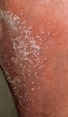 Pustular psoriasis. Note clearly defined, raised bumps on skin that are filled with pus (pustules). Skin under and around these bumps is reddish. Image from Hon Pak, MD.
Clinical severity ranges from localized palmoplantar disease to fulminant generalized eruptions associated with fever, leukocytosis, metabolic abnormalities, and life-threatening systemic complications. Recognition of the IL-36 pathway has transformed understanding of disease pathogenesis and led to development of targeted biologic therapies capable of rapidly controlling acute flares.
This review summarizes current concepts regarding the classification, pathogenesis, histopathology, diagnosis, and management of pustular psoriasis.
Pustular psoriasis encompasses several clinically distinct disorders that have the common feature of sterile pustule formation. Although it has historically been grouped with psoriasis vulgaris because of overlapping histopathologic findings, it is increasingly recognized as immunologically distinct. Unlike plaque psoriasis, which is largely driven through adaptive immune pathways involving IL-23 and IL-17, pustular psoriasis demonstrates stronger innate immune and neutrophilic inflammatory mechanisms. Mutations involving the genes IL36RN, CARD14, and AP1S3 support this distinction.
Patients appear distressed, often tachypneic, tachycardic, and febrile. The oropharyngeal mucosa may be hyperemic, and a geographic tongue or fissured tongue may be appreciated. Skin findings include a generalized or patchy erythema studded with interfollicular pustules that may have an annular or generalized/nonspecific configuration. In GPP, the skin initially becomes fiery red and tender. Constitutional signs and symptoms include headache, fever, chills, arthralgia, malaise, anorexia, and nausea. Within hours, clusters of nonfollicular superficial 2- to 3-mm pustules may appear in a diffuse pattern.
Patients with pustular psoriasis may demonstrate any of the following comorbid conditions:
-
Psoriatic arthritis
-
Metabolic syndrome
-
Cardiovascular disease
-
Depression and anxiety
Palmoplantar pustulosis (PPP) additionally demonstrates associations with smoking and thyroid disease.
Laboratory findings include the following:
-
Complete blood count (CBC) with absolute lymphopenia coinciding with polymorphonuclear leukocytosis up to 40,000/µL
-
Elevated erythrocyte sedimentation rate (ESR)
-
Elevated C-reactive protein (CRP) [3]
-
Serum chemistries - Increased plasma globulins; decreased albumin, calcium, and zinc; elevated blood urea nitrogen (BUN) and creatinine if the patient is oligemic; elevated liver enzymes if liver damage has occurred
-
Urinalysis - Positive albumin; positive casts
-
Bacterial cultures and sensitivities of pustules - Negative in the absence of secondary infection, as are Tzanck preparations and viral cultures; loss of the cutaneous barrier may result in bacteremia
On histologic evaluation, the overall architecture of the epidermis is similar to that in patients with plaque psoriasis (psoriasis vulgaris), exhibiting parakeratosis, elongation of rete ridges, and thinning of the suprapapillary epidermis. The superficial dermis shows a mononuclear infiltrate and numerous neutrophils migrating from papillary capillaries to the epidermis. Neutrophils in the epidermis can aggregate between keratinocytes, where there is also spongiosis, forming pustules known as spongiform pustules of Kogoj, a characteristic histologic feature.
There is no established standard therapy specifically for pustular psoriasis, though spesolimab comes closest. [4] Disease severity and extent of skin involvement help guide treatment. Recognition of systemic involvement, careful differentiation from mimickers, and prompt institution of effective therapy are critical. The emergence of IL-36 receptor antagonist (IL-36Ra) therapy represents a major therapeutic breakthrough and may substantially improve outcomes for patients with generalized pustular psoriasis.
General guidelines for the management of psoriasis have been developed jointly by the American Academy of Dermatology (AAD) and the National Psoriasis Foundation (NPF). [5, 6, 7, 8, 9]
Pathophysiology
GPP (historically termed von Zumbusch psoriasis) is characterized by diffuse erythema with widespread sterile pustules. It is a chronic and relapsing condition that presents with a sudden onset of rash and pustules located on nonacral skin. It also is commonly accompanied by systemic inflammation, including fever, pain, and malaise, the severity of which vary from case to case, as well as plaque psoriasis (psoriasis vulgaris). [3] However, the presence of systemic inflammation and plaque psoriasis is not necessary for diagnosis. [3, 10]
In a 2024 consensus statement, the International Psoriasis Council (IPC) defined GPP as “a systemic inflammatory disease characterized by cutaneous erythema and macroscopically visible sterile pustules.” [11] An essential diagnostic criterion in this definition was that the pustules are "not restricted to the acral region or within psoriatic plaques.”
The annular (or circinate) type is also known as subacute GPP. It tends to run a subacute or chronic course with fewer systemic manifestations. A disproportionately high number of cases are found in the pediatric population. [12]
PPP consists of recurrent sterile pustules localized to the palms and soles. Many investigators consider PPP to be distinct from classic psoriasis because of differing epidemiology, genetics, and therapeutic responses. [13] PPP is not typically associated with the life-threatening complications seen in GPP; however, possible complications include skeletal and joint disease. [3]
Acrodermatitis continua of Hallopeau (ACH) is a chronic pustular disorder involving distal digits and the nail apparatus that may result in nail destruction and osteolysis; the underlying joints and bone are spared. [3] Cases are generally refractory to treatment. Subsets of these cases are considered variants of pustular psoriasis, particularly since they are indistinguishable histologically and in early clinical presentation. [10, 14]
Mixed forms of pustular psoriasis are commonly seen in patients with pustule types and locations specific to several of the forms of pustular psoriasis described above. [10]
A juvenile or infantile type of pustular psoriasis has been described, but it is the least common form.
Additionally, several disease entities are considered by some to be variants of pustular psoriasis. These include the following:
-
Pregnancy-associated impetigo herpetiformis - Occurring predominately in the third trimester, this is a variant of acute pustular psoriasis that carries an increased risk of subsequent stillbirth or fetal abnormalities [15]
-
Sneddon-Wilkinson syndrome or subcorneal pustular dermatosis (SCPD) - This follows a relapsing and remitting course that may develop into GPP; occurring predominantly in patients middle-aged or older, it is associated with underlying malignancies (most commonly multiple myeloma and IgA monoclonal gammopathy) and pyoderma gangrenosum [16]
-
Acute generalized exanthematous pustulosis (AGEP) - Characterized by a widespread rash evolving into pustules, this is associated with a prescribed drug (eg, pristinamycin, amoxicillin, and other antibiotics [3] ) in more than 90% of cases and is considered by some a drug-induced form of pustular psoriasis [17] ; some patients develop systemic involvement, most commonly hepatic, renal, or pulmonary; AGEP is associated with IL36RN mutations similar to those found in pustular psoriasis, PPP, and ACH (see Etiology), which is not surprising in view of the clinical and immunologic similarities
Etiology
Enhanced polymorphonuclear leukocyte (PMNL) chemotaxis is much more pronounced in pustular psoriasis than in plaque psoriasis. This observation has been attributed either to an intrinsic PMNL defect or to the presence of chemoattractants in the psoriatic epidermis. Although the principal stimulus that triggers the phenomenon of massive PMNL migration from the vasculature to the epidermis is unknown, several pathways involved directly and indirectly with neutrophil chemotaxis have been investigated.
The major advance in the understanding of pustular psoriasis in the past few years has been recognition of dysregulated IL-36 signaling. IL-36 cytokines stimulate the following:
-
Keratinocyte activation
-
Chemokine production
-
Neutrophil recruitment
-
Amplification of innate immune inflammation
Normally, IL-36 signaling is regulated by IL-36Ra, encoded by IL36RN. Loss-of-function mutations result in uncontrolled neutrophilic inflammation. IL-36 receptors are expressed constitutively on dermal dendrocytes, CD4+ T cells, and macrophages; when activated, they promote maturation of monocyte-derived dermal dendrocytes and inductions of cytokines, including IL-1, IL-6, IL-23, tumor necrosis factor (TNF)-α, and interferon gamma, which promote neutrophil migration, as well as dysregulated activation of dendritic cells and T cells. [3, 18, 19]
Homozygous, compound heterozygous, and single heterozygous missense mutations in IL36RN have been associated with an increased incidence of pustular psoriasis, as well as an earlier age of onset. [3] Disease severity varies between these genotypes, with individuals who completely lack the protein having the worst prognosis. [3] These mutations predispose individuals to autosomal recessive inherited and sporadic GPP, AGEP, ACH, and, to a lesser extent, PPP. [13, 17, 20, 21]
Mutations affecting IL36RN are strongly associated with GPP, particularly early-onset disease. These mutations lead to unopposed release of inflammatory cytokines, including IL-6, IL-8, IL-1a, IL-1b, IL-23, TNF-α, and interferon gamma, which promote neutrophil activation and migration. Individuals with these mutations have a lower rate of concurrent plaque psoriasis than do patients who have pustular psoriasis without the mutation. [3]
A subset of IL-23‒responsive CD4+ T-cells, identified as type 17 helper T (Th17) cells, induce IL-17 and IL-22; these, in turn, induce production of IL-6, IL-8, and C-X-C motif chemokine 5 (CXCL5 or ENA78), which promote differentiation, activation, and migration of neutrophils and provide a positive feedback loop for Th17 cell differentiation. Significantly increased levels of IL-17 have been identified in lesional skin from patients with pustular psoriasis as compared with nonlesional skin from the same patients. [18]
IL-6 signaling has gained attention for its role in the pathogenicity of pustular psoriasis. The IL-6 receptor (IL-6R) subunit functions as both a membrane-bound receptor and a soluble receptor. This dual functionality separates it from all other known cytokine receptors, which function only as membrane-bound forms. The IL-6/IL-6R complex, together with the ubiquitously expressed gp130, activate the JAK/STAT kinase and the RAS/MEK/ERK/MAPK kinase pathways, which ultimately augment nuclear gene expression. Downstream effects of IL-6 include the following [18] :
-
Synthesis of acute-phase reactants
-
B-cell maturation
-
T-cell differentiation
-
Positive influence on Th17 cell development
-
Maturation of neutrophils from myeloid progenitors
-
Increased expression of intercellular adhesion molecule (ICAM)-1 and other endothelial adhesion molecules that enhance neutrophil migration
-
Release of proinflammatory cytokines (eg, IL-23 and IL-17) to further promote the Th17 positive feedback loop
Electron microscopic studies have shown the presence of basal keratinocyte herniations in lesions of pustular psoriasis. These are cytoplasmic processes from basal keratinocytes that protrude into the dermis through gaps in the basal lamina. These herniations are mostly clustered over collections of neutrophils in the dermis. This finding suggests an increased production of neutrophilic proteolytic enzymes in the dermis of pustular psoriasis patients.
Immunohistochemical methods have confirmed the involvement of some of these proteases and their inhibitors in the development of pustules.
Elastase is a proteolytic enzyme released by PMNLs during the process of extravasation and migration through the dermoepidermal junction. One study found an epidermal elastase inhibitor (skin-derived antileukoproteinase) expressed in psoriatic skin prior to the influx of PMNLs, which disappeared when the composition of the infiltrate changed. This finding was not confirmed by other studies.
Additional studies investigating other potential mechanisms have shown decreased natural killer (NK) cell activity in generalized pustular psoriasis. An increased incidence of human leukocyte antigen (HLA)-B27 also has been found among patients with pustular psoriasis. This haplotype is seen in psoriasis patients with peripheral arthritis, as well as in patients with ankylosing spondylitis and reactive arthritis.
Mutations in two other genes are also seen in individuals with pustular psoriasis. One gene, CARD14 (caspase-activating recruitment domain, member 14), is known to be upregulated in individuals with GPP and PPP, and this upregulation leads to increased amounts of nuclear factor (NF)-κB, IL-8, and IL-36γ. [22, 3] Mutations in the other gene, AP1S3, impair autophagy and promote inflammatory cytokine production, leading to increased amounts of IL-36α and also causing disturbances in the innate immune response through disruption of Toll-like receptor (TLR)-3. [23, 3] This is most commonly seen in individuals with GPP and ACH.
Overlap with TNF-α, IL-17, and IL-23 pathways likely explains the efficacy of biologic agents targeting these cytokines.
Triggers
Common triggers of pustular psoriasis include the following:
-
Withdrawal of systemic corticosteroids [24]
-
Infection [25]
-
Pregnancy [3]
-
Emotional stress
-
Hypocalcemia
Drugs that may act as triggers include the following:
-
Lithium
-
Hydroxychloroquine
-
Terbinafine [26]
-
Beta blockers
Paradoxic pustular eruptions have also been reported with TNF inhibitors. In many patients, the condition is idiopathic.
Epidemiology
US and international statistics
Pustular psoriasis is uncommon in the United States, accounting for only about 3% of cases of psoriasis. [27] It is rare globally as well. Prevalence figures cited for GPP include 7.46 cases per 1 million people in Japan, 1.76 per 1 million in France, and 88-124 per 1 million in the Republic of Korea. [28]
Age-, sex-, and race-related demographics
The average age of adult patients with pustular psoriasis has been reported to be in the range of 48-50 years. [29] The average age of onset of acute GPP is 31 years, and the mean ages of onset for PPP and ACH are 43.7 and 51.8 years, respectively. [13] Children aged 6 weeks to 10 years can be affected, albeit rarely. One case described generalized pustular psoriasis in a 6-week-old infant. [30] The mean age of onset of annular pustular psoriasis in pediatric populations is 6 years. [12]
In the United States, the male-to-female ratio for pustular psoriasis is 1:1. Globally, a female predominance has been reported, [31] with a female-to-male ratios of 1.5, 1.7, and 3.5, for ACH, GPP, and PPP, respectively. [13, 29, 32] PPP occurs rpedominatly in middle-aged women and has a strong association with smoking. In children, the female-to-male ratio is 3:2.
Pustular psoriasis affects all races but may be more common in some non-White ethnic groups. A study by Yan et al found a higher frequency of pustular psoriasis in Asians and Hispanics/Latinos than in Whites. [33]
Prognosis
Older patients with GPP have a poor prognosis. Death can result from sepsis or renal, hepatic, or cardiorespiratory failure during the acute erythrodermic stage. Death secondary to cardiorespiratory failure is more likely to occur in untreated patients.
Patients with a history of chronic plaque psoriasis before generalized pustular eruption tend to have a better prognosis than patients with more atypical forms of psoriasis.
PPP and ACH frequently follow chronic relapsing courses with substantial impairment in quality of life.
Children with pustular psoriasis typically have a good prognosis, as long as serious secondary infections are avoided.
There is no cure for pustular psoriasis. Recurrent flares are common, even years after diagnosis. Continued therapy and ongoing avoidance of precipitating factors are often necessary. [29] Case reports have described psoriasis flare-ups in individuals with COVID-19, including those with pustular psoriasis. [34, 35] GPP may produce recurrent life-threatening flares that necessitate hospitalization. The development of IL-36–targeted therapy may significantly improve long-term outcomes.
-
Pustular psoriasis. Note clearly defined, raised bumps on skin that are filled with pus (pustules). Skin under and around these bumps is reddish. Image from Hon Pak, MD.
-
Palmoplantar pustular psoriasis: type of pustular psoriasis appearing on palms of hands or soles of feet. Image from Hon Pak, MD.


